LYMPH NODES:
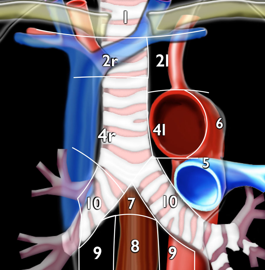
Eventually, all lymph vessels lead to lymph nodes. Realtek multifunction devices driver download for windows 10. Lymph nodes can be as small as the head of a pin, or as big as an olive. There are 400-700 lymph nodes in the body, half of which are located in the abdomen, and many are in the neck.

The primary function of lymph nodes is to filter and purify the lymph. The lymph nodes produce various types of lymphocytes. Lymphocytes destroy harmful substances within the body, and are a big part of the immune system. The lymph nodes reabsorb about 40% of the liquid content of the lymph. This makes the lymph much thicker. Because of this thickening and the filtering process, the lymph nodes offer the greatest resistance to the flow of lymph. In fact the lymph nodes offer about 15 times more resistance than the vessels themselves. Lymphatic drainage can help overcome this resistance and get the lymph flowing.
At this point we may be able to see your immediate drainage pathways, which we ‘map’ on the skin with a skin-safe marker. This is followed by the ‘wait time’, as it can take around 4 hours for the tracer to move through damaged lymphatics. Manual Lymph Drainage (MLD) This is a gentle, non-invasive manual technique that has a powerful effect on the body. Research in Australia, Europe and North America has proven its efficacy as a stand-alone treatment and in combination with other therapies. Drink PLENTY Of Fluid. Lymph is about 95% water, making water essential for its. Lymph Chains & Their Drainage Areas Lymph Nodes of the Head and Neck. Lymph Nodes at Surface; Deep Lymph Nodes; Lymph Nodes of Breast and Arm. Lymph Nodes of the Breast and Upper Limb; Thoracic Lymph Nodes. Parietal Lymph Nodes of the Thorax; Visceral Lymph Nodes of the Thorax; Lymph Nodes of the Lower Thorax; Abdominal Lymph Nodes.
EDEMA:
Each cell is nourished by the nutrients, oxygen and proteins that flow across the walls of capillaries into the interstitial fluid. There is a dynamic balance between the forces that help those nutrients to first exit the capillaries, and then get reabsorbed back into the blood stream. Proteins play a big part in this transfer because they have a tendency to draw water to themselves. This means that the proper amounts of protein on both sides of the capillary wall are vital to keep the tissues balanced. If there are too many proteins within the interstitial spaces, fluid will start to accumulate, causing edema. The lymph system’s role of removing proteins is vital to keeping edema down. If the lymph system becomes sluggish, or is damaged by surgical removal of lymph nodes, edema can develop. This type of edema is called lymphostatic edema- or a high protein edema. Lymphatic drainage can be helpful in reducing this type of edema because the cause is a reduced functioning of the lymph system.
Lymphatic Map
Other causes of edema can be a chemical imbalance in the body caused by liver disease, diabetes, or a variety of other ailments. This type of edema is called lymphodynamic edema, and requires other forms of therapy due to the fact that it is a chemical imbalance. (Kasseroller, R., Compendium of Dr. Vodder’s Manual Lymph Drainage, Haug, Heidelberg, 1998)

Lymph Node Drainage Map

this is an image of the nodes and drainage patterns of the lymphatic system
